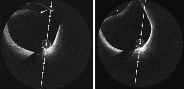
Fig. 3.
(Left-hand panel) Optical coherence tomography image of the left anterior descending artery demonstrating intimal tear (indicated by white arrow). (Right-hand panel) Optical coherence tomography image of the left anterior descending artery demonstrating intramural hematoma (asterisk) compromising the true lumen.

