
Figure 2.
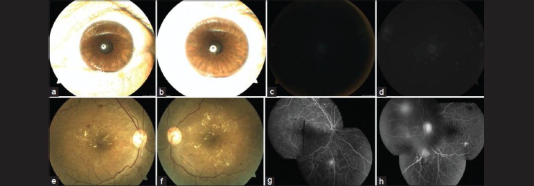
Anterior segment photographs of the right (a) and left (b) eyes of a 41-year-old male showing undilated pupils just before obtaining NMRI. The NMRI showed grade 5 quality photographs in both right (c) and left (d) eyes. Both eyes were ungradable for detection of diabetic retinopathy. Dilated fundus photographs (e and f) and fluorescein angiography photographs (g and h) of same patient as in a-d, showing grade 1 quality images. There is presence of clinically significant macular edema and neovascularization elsewhere on the retina in both eyes, suggestive of proliferative diabetic retinopathy