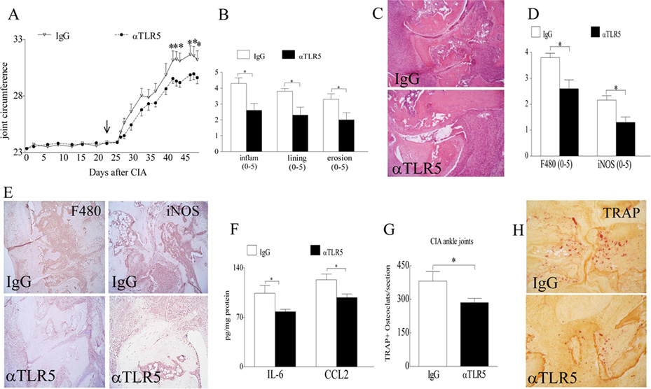
Figure 8. Anti-TLR5 antibody treatment alleviates CIA joint swelling and bone resorption.
A. Changes in joint circumference were recorded for CIA mice that were treated i.p. with IgG or anti-TLR5 Ab (100 µg/mouse) on days 23, 27, 30, 34, 37, 41, 44 and 48 and mice were sacrificed on day 49 post induction, n=6 mice (12 ankles). B. Effect of anti-TLR5 Ab treatment on inflammation, lining thickness, and bone erosion was scored on a 0–5 scale, n=6. C. Is the representative ankle H&E staining (original magnification × 200) of Fig. B. D. Synovial tissues from CIA mice treated with IgG or anti-TLR5 antibody were harvested on day 49 and immunostained with anti-F480 (1:100 dilution) or iNOS Abs (1:200 dilution) (original magnification × 200). Joint myeloid cells and iNOS+ M1 macrophages staining were quantified on a 0–5 scale, n=6. E. Is the representative F480 and iNOS immunostaining (original magnification × 200) of Fig. D. F. Changes in IL-6 and CCL2 protein levels in ankle homogenates from CIA mice treated with IgG control or anti-TLR5 antibody were determined by ELISA, n=6. G. Number of TRAP+ cells were counted per section in CIA mice treated with IgG or anti-TLR5 Ab, n=6. H. Is the representative ankle TRAP staining (original magnification × 200) of Fig. G. Values are mean ± SE. *p < 0.05.

