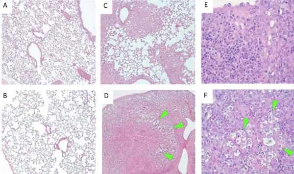
Figure 3. Uncontrolled C. neoformans expansion and severe lung pathology develop in H99γ-infected STAT1 KO mice by day 10 post-infection.
129S6/SvEv (WT) and STAT1 KO mice were given an intranasal inoculation with 1 × 10⁴ CFU of C. neoformans strain H99γ or left uninfected. Lungs of uninfected (A-B) and H99γ-infected (C-F), WT (upper row) and STAT1 KO mice (lower row) were collected on day 10 post-inoculation, processed and analyzed using light microscope. Deletion of STAT1 that did not affect morphology of uninfected lungs has resulted in the development of severe lung pathology and massive expansion of fungus by day 10 post-inoculation. Note that the infection and inflammatory response are contained within portions of the lungs in the WT mice (C), while virtually all lungs are infected and consolidated in STAT1 KO mice (D). High power images show minimal presence of cryptococcal organisms and predominantly mononuclear cell infiltrates with lymphoid and macrophage-type morphologies within infected lungs areas in the WT mice (E), contrasting with granulocyte-enriched mixed cellular infiltrate surrounding clusters of proliferating cryptococci (green arrows) and in the lungs of STAT1 KO mice (F). Histological slides were stained with H&E and mucicarmine, and images were taken at 10X (A-D) and 40X (E-F) objective power. Images are representative of images derived from 2 experiments using 3 mice per group.

