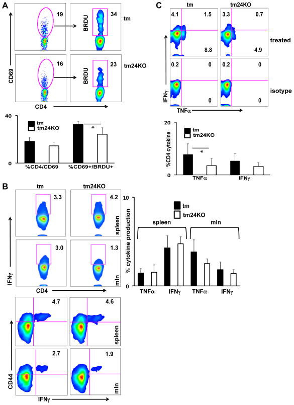
Figure 2. Decreased T cell activation, proliferation, and cytokine production in tm24KO mice.
Tm and tm24KO mice were subjected to flow cytometric analysis. CD4+ single-positive populations were gated on. (A) Splenocytes from 6 month tm or tm24KO mice were surface stained for CD69+ and intracellular stained for BrdU after 3 days of BrdU water treatment. Data of one representative pair of mice from 4 pairs of mice is shown. Graph depicts statistical significance for CD4/CD69+ and CD4/CD69/BrdU+ cells, (*=p<0.05) (B) Splenocytes or mesenteric lymph node cells were incubated with PMA/Ionomycin in the presence of brefeldin A for 4–6 hours. Intracellular stain of IFN-γ (shown) and TNF-α were performed. Top flow panels show CD4-specific IFN-γ production. Bottom panels are gated on CD4+ single-positive cells and assessed for CD44/IFN-γ double positive populations. Data shown are one pair of mice, representative of 4 pairs of mice. Graph depicts statistical significance for CD4/TNF-α+ or CD4/IFN-γ+ splenocytes and mesenteric lymph node cells. (C) Peripheral blood mononuclear cells were incubated with PMA/Ionomycin in the presence of brefeldin A for 4 -6 hours. Intracellular stain of IFN-γ (shown) and TNF-α were performed. Data of one representative pair of mice from 5 pairs of mice is shown. Graph depicts statistical significance for CD4/TNF-α+ or CD4/IFN-γ+ PBMCs, (*=p<0.05).

