
Fig 1.
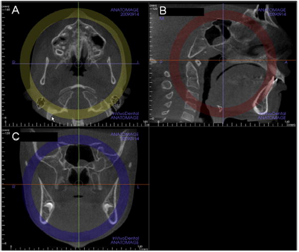
Standardization of head position in the A, axial; B, sagittal, and C, coronal planes to allow consistent assessments of the midpalatal suture. Note that in B, the sagittal view, the orange line that indicates the position of the axial plane view is positioned through the center of the superoinferior dimension of the hard palate (Invivo5).