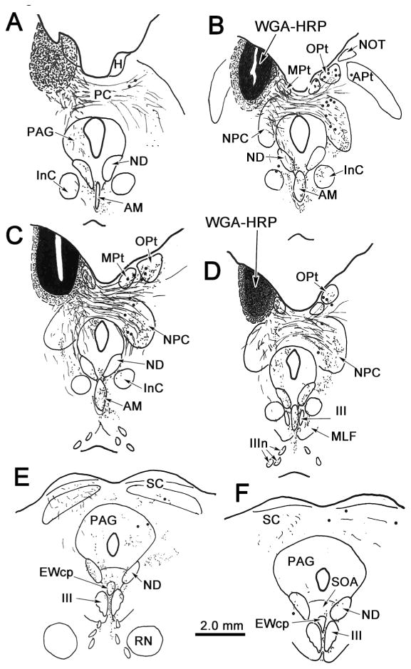
Figure 2.

Distribution of labeled cells and terminals in the midbrain following an injection of WGA-HRP into the left olivary pretectal nucleus (OPt). In this and all subsequent chartings, sections are arranged from rostral (A) to caudal (F), retrogradely labeled cells are indicated by dots, anterogradely labeled terminals are indicated by fine stipple and fine lines represent labeled axon fibers. Injection site and tracer spread area are indicated by black and shaded areas, respectively (A-D). The injection site was centered in OPt and spread slightly into adjacent parts of the medial pretectal nucleus (MPt), the nucleus of the optic tract (NOT), and the anterior pretectal nucleus (APt). Terminals are present bilaterally in the anteromedian nucleus (AM) (A-D), and to a lesser extent in the centrally projecting Edinger-Westphal nucleus (EWcp), supraoculomotor area (SOA) and between the oculomotor nuclei (III). Terminals are also present in the ipsilateral periaqueductal gray (PAG)(A-E), both nuclei of Darkschewitsch (ND)(A-F), and contralaterally in OPt (B-D), MPt (B-D), the nucleus of the posterior commissure (NPC)(B-D) and the interstitial nucleus of Cajal (INC)(B&C). Labeled cells are present in contralateral OPt, MPt, APt and NPC (B-D).
