
Figure 5.
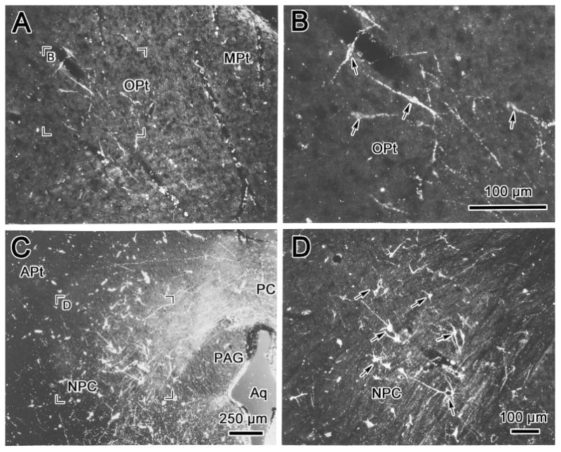
Darkfield photomicrographs showing retrogradely labeled cells in the olivary pretectal nucleus (OPt)(A, B), and in the nucleus of the posterior commissure (NPC)(C, D) following a WGA-HRP injection into the anteromedian nucleus (AM) like that shown in figure 4. Examples of the labeled cells are indicated by arrows (B,D&F). The region selected for the high magnification views (B&D) is indicated in the low magnification views (A&C). Scale in A=D.