
Figure 7.
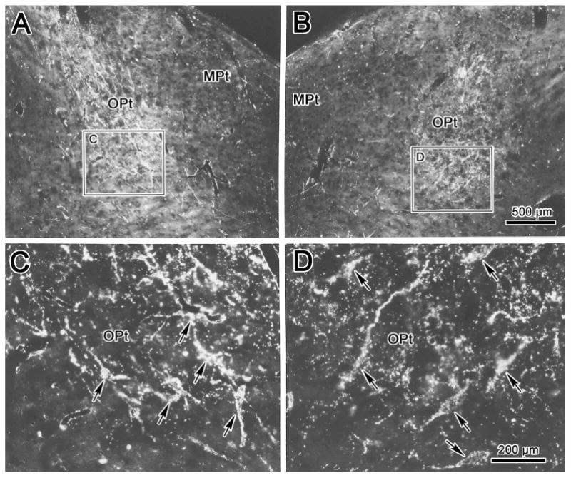
Darkfield photomicrographs showing labeling in the olivary pretectal nucleus (OPt) following combined injections of WGA-HRP into the left vitreous chamber and into the anteromedian nucleus. Lower magnification (A&B) and higher magnification (C&D) views from regions indicated by boxes reveal distribution of label in the left (A&C) and right (B&D) OPt. Premotor neuons retrogradely labeled from the AM nucleus overlaped with anterogradely labeled retinal terminals. Examples of these labeled cells are indicated by arrows. Scale in A=B. C=D.