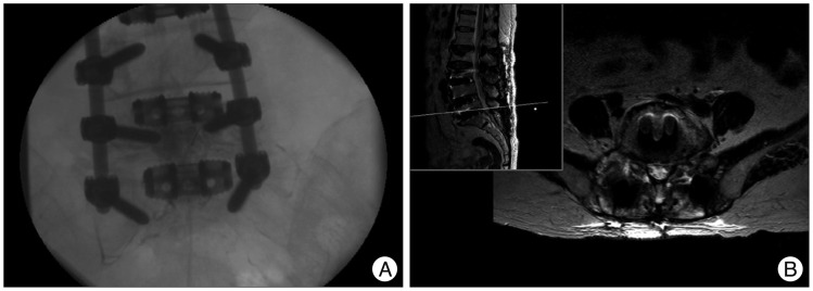
Fig. 4.
Images obtained during percutaneous epidural neuroplasty (A). It was performed especially focused on right L5/S1 foramen. MRI obtained after the third operation (B), showing the same level as Fig. 3C, revealed facetectomy and decompression state at right L5/S1 extraforaminal region.

