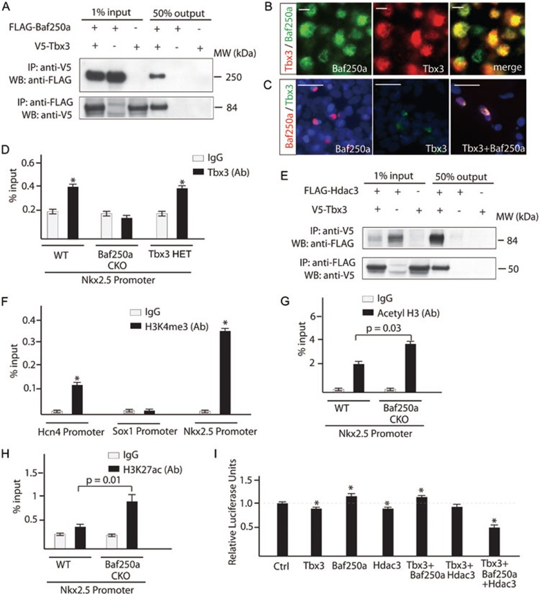
Figure 3.
Baf250a, Tbx3, and HDAC3 coordinately repressed the expression of Nkx2.5 in the SAN. (A) Baf250a directly interacts with Tbx3. Baf250a or Tbx3 was immunoprecipitated from nuclear extracts of HEK 293 cells expressing FLAG-tagged Baf250a, V5-tagged Tbx3, or both proteins. Association with Tbx3 or with Baf250a was detected by western blotting with V5- or FLAG-specific antibody. (B) Co-localization of Baf250a and Tbx3 in the pacemaker cells of SAN. Scale bar, 5 μm. Magnification: 400×. (C) Either Baf250a or Tbx3 alone primarily localized in the cytosol of 293T cells. Co-expression of Baf250a and Tbx3 led to exclusive nuclear localization of both proteins. Blue: DAPI. Scale bar, 25μm. (D) Tbx3 binding to promoter of Nkx2.5 required Baf250a. Binding of Tbx3 to the promoter of Nkx2.5 in the SAN of wild-type (WT), Baf250a CKO mice and Tbx3 HET mice. WT: wild type as determined by ChIP-qPCR (n = 3 in each group). (E) Tbx3 directly interacts with HDAC3. Reciprocal Co-IP was similarly performed as described in A. (F) ChIP-qPCR showed the distribution of H3K4me3 in the proximal promoters of Hcn4, Sox1, and Nkx2.5 (n = 3 in each group). *P < 0.05. (G) Increased acetylation of histone 3 in the promoter region of Nkx2.5 at 20 h after tamoxifen induction in the SAN of Baf250a CKO mice as determined by ChIP-qPCR (n = 3 in each group). (H) ChIP-qPCR evaluated the distribution of H3K27ac in the proximal promoter of Nkx2.5 in Baf250 CKO mice. (I) Baf250a, Tbx3, and HDAC3 coordinately repressed the promoter activity of Nkx2.5 in a luciferase reporter assay (n = 3 in each group). *P < 0.05.

