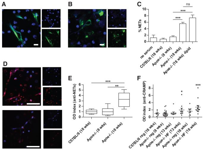
Figure 2. Apoe−/− mice demonstrate enhanced neutrophil extracellular trap (NET) formation and develop autoantibodies to NETs.
A and B, Representative immunofluorescence staining of nonpermeabilized Apoe−/− neutrophils for citrullinated histone H3 (H3-Cit; A) and MPO (B). DNA is stained blue, and the indicated protein green. Scale bars=10 μm. C, Bone marrow neutrophils were isolated from 8-week-old Apoe−/− mice. Neutrophils were incubated in the presence of 10% serum from the indicated mice for 4 hours (n=5 mice per group). ***P<0.001. D, Apoe−/− NETs were fixed and incubated with 1% serum from 18-week-old Apoe−/− mice (top) or 8-week-old Apoe−/− mice (bottom). Detection of bound antibodies was with Texas-Red-conjugated anti-immunoglobulin G; DNA is stained blue. Scale bars=50 μm. E, NET proteins were prepared as described in Methods and used to coat plates for the enzyme-linked immunosorbent assay (ELISA). Optical density (OD) index normalizes data to the average value for C57BL/6 mice. Box-and-whisker plots show data for 8 mice per group, with boxes representing the median, 25th percentile, and 75th percentile; whiskers delineate the minimum and maximum values. **P<0.01; ***P<0.001. F, An ELISA for anti-cathelicidin-related antimicrobial peptide (CRAMP) was performed as described in Methods. Some Apoe−/− mice were placed on high-fat chow (HF) beginning at 8 weeks of age; others remained on regular chow (reg). OD index normalizes data to the average value for control mice. Mean and SEM are plotted, with n≥8 per group. *P<0.05 and ***P<0.001 when compared with C57BL/6 control mice.

