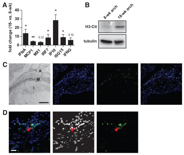
Figure 3. Interferon expression and histone citrullination are upregulated in atherosclerotic lesions.
A, Apoe−/− mice were placed on high-fat chow beginning at 8 weeks of age. RNA was isolated from aortic arches of 8- and 18-week-old Apoe−/− mice. Fold change in gene expression was calculated for 18-week-old mice, relative to 8-week-old mice (n=5). Mean and SEM are plotted. *P<0.05, **P<0.01, and ***P<0.001; P values that did not reach significance are indicated. B, Protein was prepared from aortic arches of the indicated Apoe−/− mice. Protein from 5 mice per group was pooled and 20 μg of total protein was resolved by sodium dodecyl sulfate-polyacrylamide gel electrophoresis before Western blotting with the indicated antibodies. C, Low magnification view of an atherosclerotic lesion with a phase-contrast image showing intima (I), media (M), and adventitia (A). DNA is stained blue, and MPO is stained green, with an overlay to the far right. Scale bar=100 μm. D, A higher magnification view of the media/adventitia interface shows an MPO-positive cell in more detail. Extracellular MPO (green) juxtaposed with decondensed DNA (blue) is seen (red arrowhead); in the middle, the DNA channel is shown in grayscale to improve contrast. Scale bar=20 μm.

