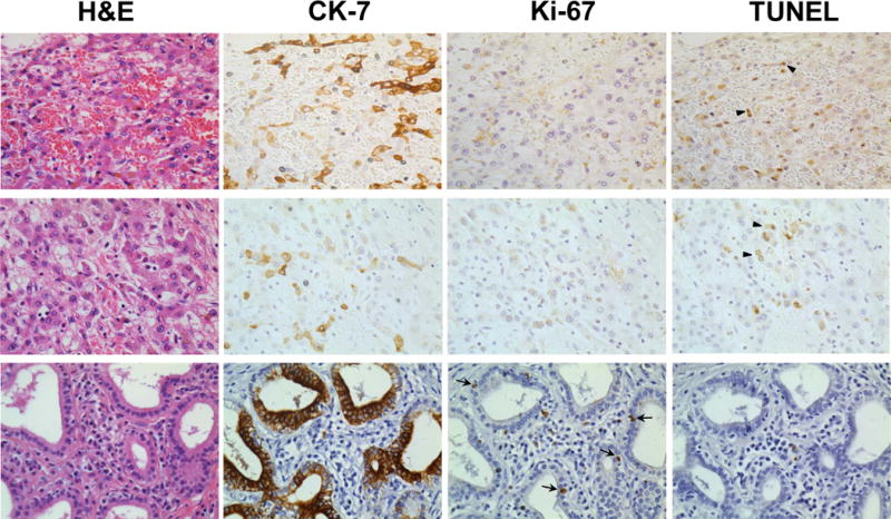
Figure 2. Immunohistochemistry for proliferating and apoptotic CCA cells.

Two representative patients with partial pathologic responses (Patient 2, top row and Patient 5, middle row) are shown. A non-treated, surgically resected CCA is shown for comparison (bottom row)._Serial sections for H&E stain, CK-7 stain for CCA cells, Ki-67 stain for proliferation, and TUNEL staining for apoptosis are indicated and performed as described in Methods. Arrows indicate dual positive TUNEL+/CK-7+ cells. 40× magnification shown.
