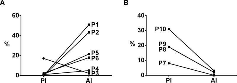
Figure 3. Proliferative and apoptotic index of hilar CCAs following neoadjuvant therapy.

Patients hilar CCAs (n=6) following neoadjuvant therapy and transplantation (A) underwent IHC for CK-7, Ki-67, and TUNEL staining as shown in Fig. 2 and as described in Methods. Proliferative index (PI) and apoptotic indexes (AI) were calculated as the percentage of Ki-67 positive CK-7 cells and percentage of TUNEL positive CK-7 cells, respectively. Patients who underwent resection only (no neoadjuvant therapy, n=4) are shown for comparison (B).
