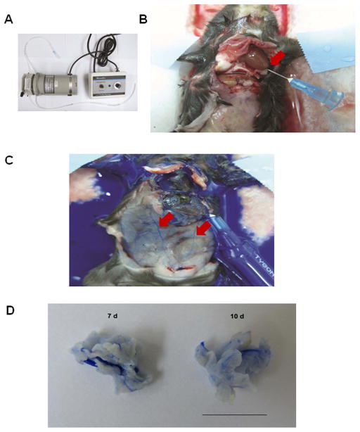
Figure 1. Perfusion of peripheral blood circulation and xenografted tumors with heparin and Microfil.
(A) Tubing and pump (pump speed, 2 mL/min). (B) Catherization in left ventricle, for exsanguination with 0.1% heparinized solution. The right atrium is nicked when the catheter is inserted (arrow). (C) Microfil perfusion through the same catheter after heparin treatment. Arrows point to Microfil perfused in peripheral blood vessels. (D) Macroscopic appearance of Microfil-perfused LLC carcinomas derived from a bolus injection of 1 × 10⁶ tumor cells in the mouse flanks at 7 d (left) and 10 d (right). Bar, 10 mm.

