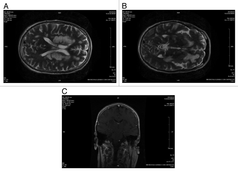
Figure 2. AxialT2encephalic magnetic resonance (A and B), coronal T1 post Gd (C) sequences performed about 1 mo after clinical onset of symptoms and after therapy: supratentorial and infratentorial demyelinating lesions were unchanged with overall dimensions slightly reduced and negative enhancement.

An official website of the United States government
Here's how you know
Official websites use .gov
A
.gov website belongs to an official
government organization in the United States.
Secure .gov websites use HTTPS
A lock (
) or https:// means you've safely
connected to the .gov website. Share sensitive
information only on official, secure websites.
