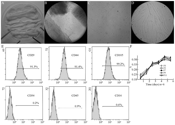
Figure 1.
Passage of hUC-MSCs in Wharton’s Jelly. (A) Wharton’s Jelly tissues without umbilical artery or vein. (B) Cells crawled out of cultured Wharton’s Jelly 2 days after culture (magnification, ×100). (C) Fusiform shape cells adhered to the bottle 5 days after culture (magnification, ×100). (D) Typical hUC-MSCs could be observed 7 days after culture (magnification, ×400). (E) The surface marker of human MSCs in Wharton’s Jelly of the human umbilical cord expressed CD44 (91.4%), CD29 (91.3%), CD105 (99.2%), but not CD34 (0.2%), CD45 (0.9%) or CD14 (0.6%). (F) Growth curves of human umbilical cord derived MSCs at 3, 5, 7, 9 and 11 passages. hUC-MSCs, human umbilical cord mesenchymal stem cells; CD, cluster of differentiation.

