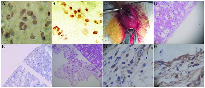
Figure 5.
5-bromo-2′-deoxyuridine (BrdU) staining of islet-like cells (magnification, ×250). (A) For cells without BrdU marking, there was no brown staining in the cytoplasm. (B) Light brown staining for BrdU-marked cells. (C) The induced islet-like cell clusters were transplanted into the renal capsule in rats. (D) The HE staining of normal rat kidney showed integral renal capsule edge, with clear morphology and cell structure. (E) The immunohistochemistry of normal rat kidney showed that the insulin and BrdU staining of the renal capsule were negative. (F) Following transplantation with induced islet-like cells, the HE staining of the renal cells showed that there were a large number of surviving cells between the renal capsule and renal cortex, with fracture of the renal capsule. (G) The kidney immunohistochemical staining showed insulin-positive cells between the renal capsule and renal cortex. (H) The immunohistochemical staining indicated BrdU-positive cells between the renal capsule and renal cortex, with brown nuclei. BrdU, bromodeoxyuridine; HE, hematoxylin and eosin

