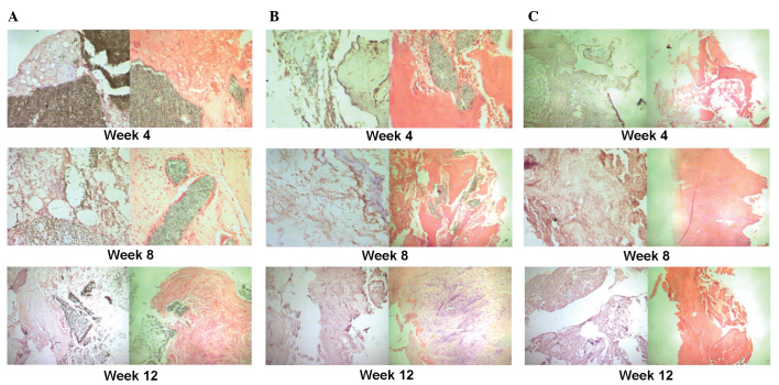
Figure 3.
Immunohistochemical staining of osteocalcin and hematoxylin and eosin staining at different time-points (4, 8 and 12 weeks) following implantation of the scaffolds. (A) Group I (pure β-TCP). At 4 weeks following implantation, new formation of cartilage and bone was observed at the edges of the scaffold (magnification, ×200). The expression of newly formed cartilage and bone increased at 8 weeks following implanation and a number of osteogenic cells arranged in a spiral were observed (magnification, ×200). At 12 weeks following implantation there was an even higher expression of newly formed cartilage and bone at the edges of the scaffold cells and the osteogenic cells were arranged in a spiral; however, the majority of the β-TCP remained detectable (magnification, ×100). (B) Group II (β-TCP/PRP). At 4 weeks following implantation, new formation of cartilage and bone was observed at the edges of the scaffold and the scaffold began to degrade (magnification, ×200). The expression of newly formed cartilage and bone increased at 8 weeks following implanation and the scaffold was further degraded and absorbed (magnification, ×100). This continued at 12 weeks following impantation where the formation of capillary vessels was also detected (magnification, ×200). (C) Group III (autogenic ilium). At 4 weeks following implantation, the new formation of cartilage, bone and capillary vessels was observed. This increased at 8 and 12 weeks following implantation with much mature bone tissue observed at 12 weeks following implantation (magnification, ×100).

