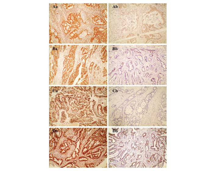
Figure 1.
Immunohistochemical staining showing normal and aberrant MMRP expression (magnification, ×100). (A) hMLH1, (B) hMSH2, (C) hPMS2 and (D) hMSH6. (Aa-Da) Normal immunohistochemical staining of (Aa) hMLH1, (Ba) hMSH2, (Ca) hPMS2 and (Da) hMSH6. Normal nuclear staining of the MMRPs can be observed not only in stromal cells, but also in epithelial tumor cells, showing a brownish accumulation of dye in the nucleus. (Ab-Db) Aberrant staining of (Aa) hMLH1, (Ba) hMSH2, (Ca) hPMS2 and (Da) hMSH6. Aberrant nuclear staining of the MMRPs can only be observed in stromal cells, not in epithelial tumor cells. MMRP, mismatch repair protein; hMLH1, human mutL homolog 1; hMSH, human mutS homolog; hPMS2, human postmeiotic segregation increased 2.

