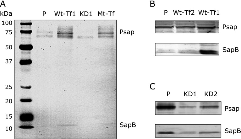
Fig. 1.
Protein measurement by western blot analysis using monoclonal anti-sapB IgG. (A) Prosaposin (Psap) and saposin B (SapB) proteins increase in Psap transfected human (Wt-Tf1) HepG2 cells and SapB domain mutated (Mt-Tf) HepG2 cells, and these proteins decrease in Psap knockdown (KD1) HepG2 cells as compared to those in parental (P) HepG2 cells. (B and C) Psap and SapB increase in Psap transfected (Wt-Tf1 and Wt-Tf2) HepG2 cells and these proteins decrease in Psap knockdown (KD1 and KD2) HepG2 cells as compared to those in parental (P) HepG2 cells.

