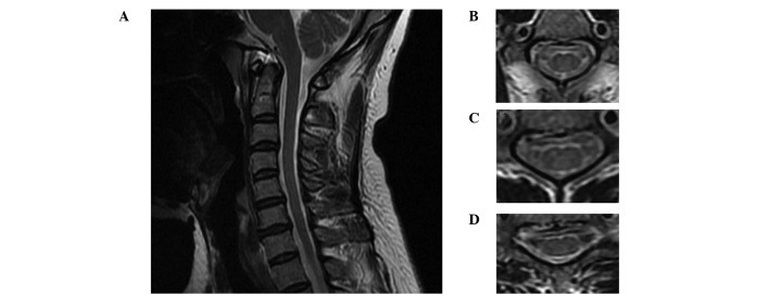
Figure 2.
MRI scans of the spinal cord. (A) Sagittal T2-weighted MRI scan showing the normal appearance of the posterior column, despite a number of disc osteophyte complexes with mild central canal stenosis. Axial T2-weighted MRI scans at the (B) C2, (C) C4 and (D) C6 levels. MRI, magnetic resonance imaging.

