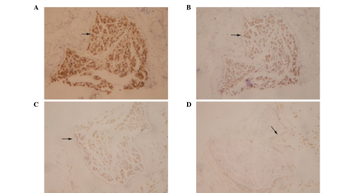
Figure 5.
Immunohistochemical analyses of the sural nerve revealed (A) S-100 strongly positive immunoreactivity (arrow), (B) NSE moderately positive immunoreactivity (arrow; abundant in the cell bodies), (C) NF mildly positive immunoreactivity (arrow) and (D) EMA mildly positive immunoreactivity. NSE, neuron-specific enolase; NF, neurofilament; EMA, epithelial membrane antigen.

