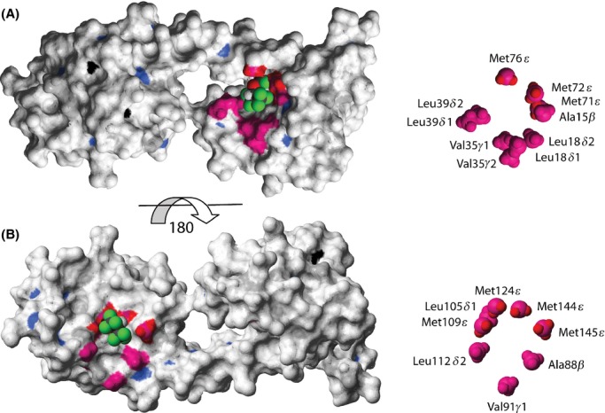
Figure 2.

Structural identification of the sevoflurane (SF)-binding sites to (Ca2+)4-CaM. Surface representation of (Ca2+)4-CaM (pdb id 1X02) (Kainosho et al. 2006) color coded accordingly: amide atoms with Δδ(1H,15N)>50 Hz in blue, methyl atoms with Δδ(1H,13C)>50 Hz in magenta, Met Hε protons with NOEs to SF in red and Ca2+ ions in black. SF, docked using Glide (Schrödinger, Inc.), is shown in CPK representation. The N-terminal binding site for SF to (Ca2+)4-CaM is shown in (A); a view of the C-terminal binding site is obtained by turning the structure 180 degrees (B). A close-up of the methyl groups of each binding interface with assignments is shown, to the right.
