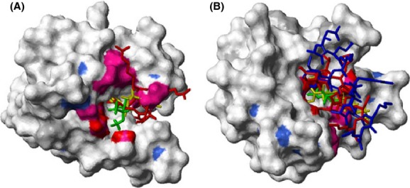
Figure 6.

Superimposed structures of (Ca2+)4-CaM binding sevoflurane (SF), W-7, and ion channel domains. Surface representation of (Ca2+)4-CaM (pdb id 1X02) (Kainosho et al. 2006) N-terminal domain (A, residues 13–80 for clarity) and C-terminal domain (B, residues 81–148). For color coding scheme, see Figure 2. Ligands are shown in stick representation with SF (docked using Glide, Schrödinger, Inc.) in green, W-7 in yellow, skeletal muscle isoform ryanodine receptor (RYR1) peptide representing (residues Arg3629–Met3638 and Ala3618–Lys3626 interacting with the N- and C-terminal domain, respectively) in red and human NaV1.5 DIII-IV linker, residues Asn1489–Lys1500, in blue.
