
Figure 5.
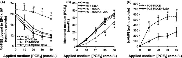
PGT regulates EP4 receptor desensitization in response to exogenous PGE2. Wild-type MDCK (“WT”) and PGT-GFP-MDCK (“PGT-MDCK”) cells were transiently transfected with the EP4-expressing cDNA, and 24 h later were exposed to pharmacological concentrations of exogenous PGE2 (0–50 nmol/L, “applied medium [PGE2]”) in the presence or absence of 5 μmol/L T26A. After 10 min, media were collected for determining residual extracellular PGE2 concentrations by ELISA, and cells were harvested for quantifying the amount of cell surface EP4 receptor using a binding assay. (A) Cell surface EP4 receptor. Exogenous PGE2 caused a reduction in cell surface EP4 (i.e., desensitization) in wild-type MDCK cells (open circles); T26A had no effect on this desensitization (closed circles). When PGT was expressed, it abrogated the ability of PGE2 to cause EP4 desensitization (open triangles), an effect that was reversed by the PGT inhibitor T26A (closed triangles). (B) Media [PGE2] at the conclusion of 10 min exposure to cell monolayers. PGT reduced the concentration of PGE2 in the media (open triangles) compared to the lack of change in wild-type cells (open circles) or wild- type cells exposed to T26A (closed circles). T26A reversed the ability of PGT to lower media [PGE2] (closed triangles). (C) Intracellular cAMP accumulation. PGT-GFP-MDCK cells (“PGT-MDCK”) were transfected with EP4 as above, treated with 250 μmol/L IBMX for 18 h, and exposed to exogenous PGE2 ± T26A for 10 min at various doses as above. Cells expressing uninhibited PGT (absent T26A, open triangles) exhibited a robust cAMP dose-response to PGE2, whereas PGT-MDCK cells in which PGT was inhibited by T26A (open triangles) exhibited a significantly blunted cAMP response to exogenous PGE2. Values are mean ± SEM (n = 3 for each experiment). *P < 0.05.