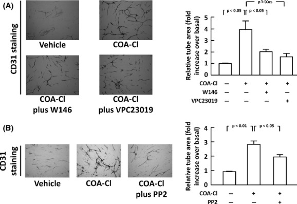
Figure 9.

Promotion of tube formation activity by COA-Cl in HUVEC and inhibitory effects of S1P1 antagonists and a Src tyrosine kinase. The results of tube formation analyses are shown. (A) HUVEC were cocultured with normal human fibroblasts for 10 days in the presence or absence of COA-Cl (100 μmol/L), with/without W146 (20 μmol/L) or VPC23019 (5 μmol/L), as indicated. Endothelial tube formation was then identified as CD31-positive signals in microphotographs, which were subsequently quantified in the graph. The right half of the figure summarizes the results derived from four independent assays, representative images of which are shown in the left half. (B) Certain cells were treated with PP2 (2.5 μmol/L). In both panels, scale bars correspond to 500 micrometer (n = 4).
