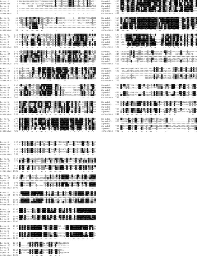
Figure 6.
Comparison of Pgp protein sequence in human, Drosophila, and locust. Multiple sequence alignment of the human Pgp (Hs-mdr1), the Dm Mdr65 (Dm-mdr65), and the three Sg Pgp/Mdr65 orthologs (Sg-mdr1-3). The protein sequence alignment was performed by Clustal Omega analysis, available from the European Bioinformatics Institute (Goujon et al. 2010; Sievers et al. 2011). All parameters were set at default values. The BoxShade algorithm (http://www.ch.embnet.org/software/BOX_form.html) was used for analysis of identical and similar amino acid residues. Highly conserved residues occurring in at least 60% of the sequences are highlighted in black. Residues similar in chemical nature occurring in at least 60% of the sequences are highlighted in gray. The consensus line includes the following symbols: “*” representing a particular position at which an amino acid is conserved in all sequences, “.” representing a position for which at least 60% of the amino acid residues are identical or similar. (The Sg-mdr1-encoding transcript sequence was only partially retrieved from the available Schistocerca gregaria transcriptome database.)

