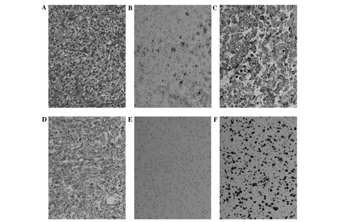
Figure 2.
Histological analysis of the mass demonstrated renal cell carcioma metastasis. (A) Hematoxylin and eosin staining identified clear cell differentiation. Immunostaining revealed (B) some positivity for cluster of differentiation 10, in addition to (C) strong positivity for pan-cytokeratin (CK). The tumor was (D) positive for vimentin and (E) negative for CK7. (F) The Mib-1 proliferation index was high and in certain areas increased to ≤70%. Magnification, ×200.

