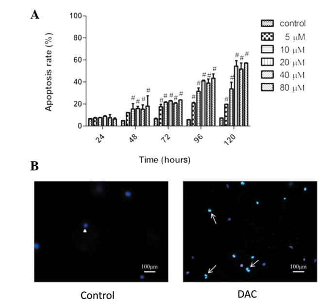
Figure 3.

DAC induces apoptosis of TFK-1 cells. (A) Cells were incubated with various concentrations of DAC for the time periods indicated. Apoptosis was measured by Annexin V and PI double-staining. #P<0.01, vs. control. (B) DAC was found to induce morphological changes of apoptosis in TFK-1 cells. Following treatment with 25 μM DAC for 120 h, cells were loaded with Hoechst 33342/PI and then observed using a fluorescence microscope. Normal cells (headed arrow) and apoptotic cells (short arrow) were identified (magnification, ×100). DAC, decitabine; PI, propidium iodide.
