
Figure 1.
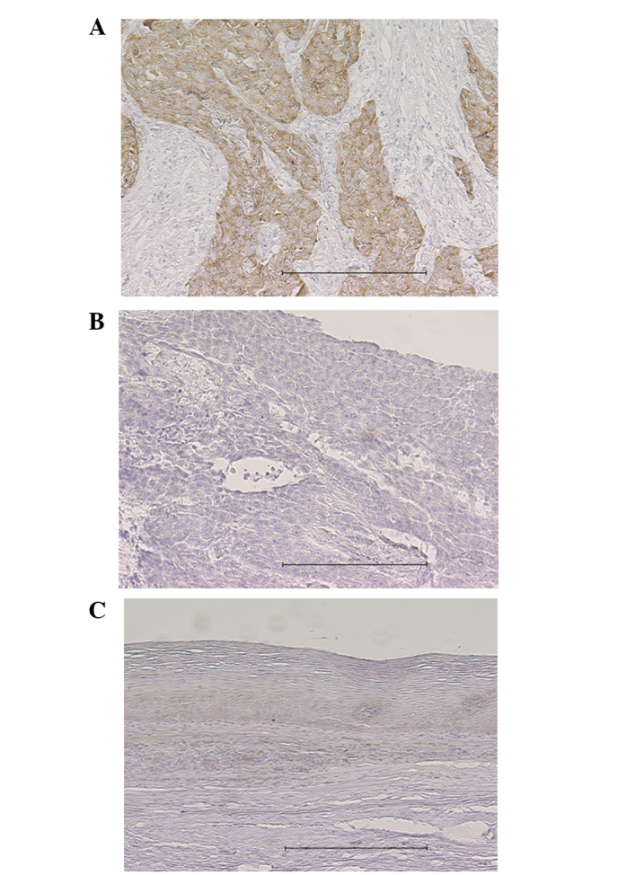
Representative images of IMP-3 expression, as determined by immunohistochemical staining. (A) IMP-3-positive esophageal squamous cell carcinoma exhibiting staining mainly in the cytoplasm of the tumor cells. (B) IMP-3-negative esophageal squamous cell carcinoma exhibiting almost no staining of the tumor cells. (C) Normal squamous epithelium negative for IMP-3. The black scale bar represents 250 μM. IMP3, insulin-like growth factor-II mRNA-binding protein-3.