
FIG. 3.
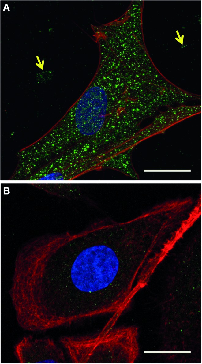
Immunofluorescent confocal microscopy showing endogenous C12orf29 expression in mandible osteoblasts (mOB) and PC3 cells. Cells were labeled with anti-C12orf29 antibody and the expression visualized using an Alexa-Fluor 488-conjugated secondary antibody shown here in green. The cell nuclei are stained with DAPI (blue) and cytoskeleton is stained with Rhodamine Phallioidin (red). Scale bars=20 μm. (A) A mOB cell showing strong C12orf29 staining throughout the cytosol and also in patches in the extracellular space (arrows). (B) A PC3 cell shows a faint but discernible C12orf29 staining within the cell. Color images available online at www.liebertpub.com/tec