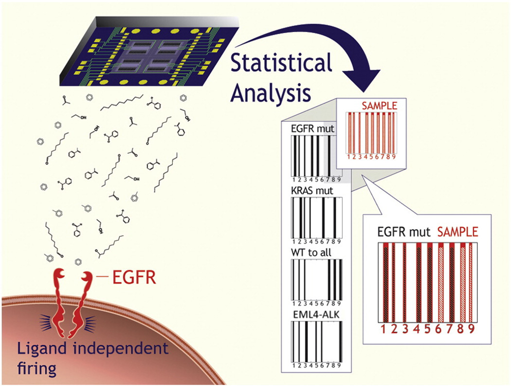
Figure 1.
Schematic representation of the oncogene identification in an unknown sample. The nanomaterial-based sensor array is exposed to the cell-line headspace, and the VOC fingerprint is obtained through DFA analysis, using three primary, general DFA models (cf. Figure 2) and, if necessary, six secondary, specific DFA models (cf. Figure 3). The oncogene is identified through comparison with the expected DFA classifications for the EGFRmut, KRASmut and EML4-ALK.

