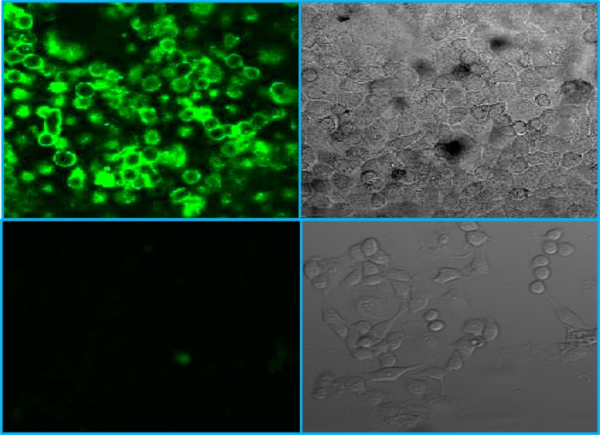
Figure 4.

Confocal microscopic fluorescence image (top left) and bright field image (top right) of HeLa cells incubated with dye-conjugated BiOI NPs. Also shown are the fluorescence image (bottom left) and the bright field image (bottom right) of untreated HeLa cells as a negative control.
