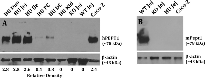
Figure 3.
Immunoblots of hPEPT1 protein in the small intestine, large intestine, and kidney of wildtype (WT = mPept1+/+), Pept1 knockout (KO = mPept1–/–), and humanized PEPT1 (HU = mPept1–/–/hPEPT1+/–) mice (A), and mPEPT1 protein in the jejunum of the same genotypes (B). Protein samples were separated by 10% SDS-PAGE, transferred onto PVDF membranes, and incubated for 1.5 h with rabbit antihuman hPEPT111 (1:3000) or antimouse mPEPT112 (1:5000) antiserum, and a mouse monoclonal antibody for β-actin (1:1000). The membranes were washed three times with TBST and then incubated for 1 h with an appropriate secondary antibody of IgG conjugated to horseradish peroxidase (1:3000). Caco-2 cells served as positive and negative controls, respectively, for hPEPT1 and mPEPT1. Duo represents the duodenum, Jej the jejunum, Ile the ileum, PC the proximal colon, DC the distal colon, and Kid the kidney.

