
Figure 7. Osteoblast alignment based on distance from crack.
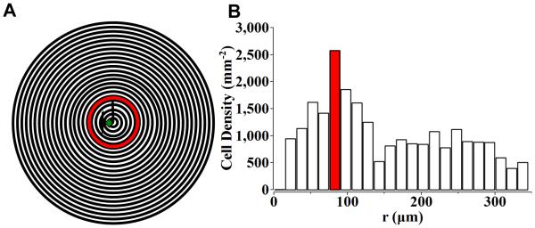
The micrograph was partitioned into a central circle of 15 μm in radius and 15 μm wide outer rings (A). The number of OBs with aligned tendency (θ<45°) in the center circle and outer rings (15 μm wide) were counted and normalized by the area respectively (B). Most cells with aligned tendency (θ<45°) were found in the annulus 75 to 90 μm from the indentation center (B), which is around the tip of the radial cracks from Vickers indentation (A).