
Fig 3. Structural studies of neuroblastoma-associated gangliosidc.
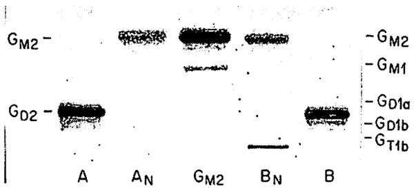
Reaction products (lanes AN and BN) of neuroblastoma tissue ganglioside (lane A) and of plasma ganglioside (lane B) had mobility identical with that of standard GM2 ganglioside, isolated from Tay-Sachs disease brain tissue (centre lane). Migration of standard brain gangliosides indicated on right.