
Figure 3. Role of IL-17a in aortic stiffening.
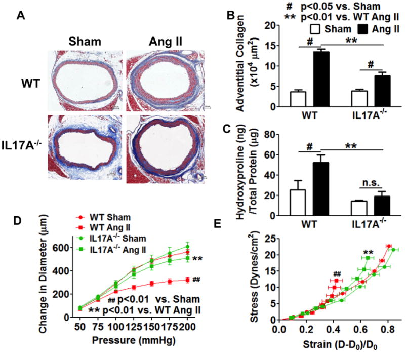
IL-17a deficient mice were infused with chronic angiotensin II (490ng/kg/min) for 14 days. A and B, the thoracic aortas were fix-perfused for Masson’s trichrome staining and quantified for adventitia collagen staining. Scale bar indicates 100 μm. C, segments of the thoracic aorta were digested in 6N HCl at 105 °C for 48 hours and measured for hydroxyproline concentration. Data analyzed using two-way ANOVA. D and E, compliance curves and stress-strain curves for IL-17a−/− mice. Ang II, angiotensin II. Data analyzed using one-way ANOVA with repeated measures, n=6–8.