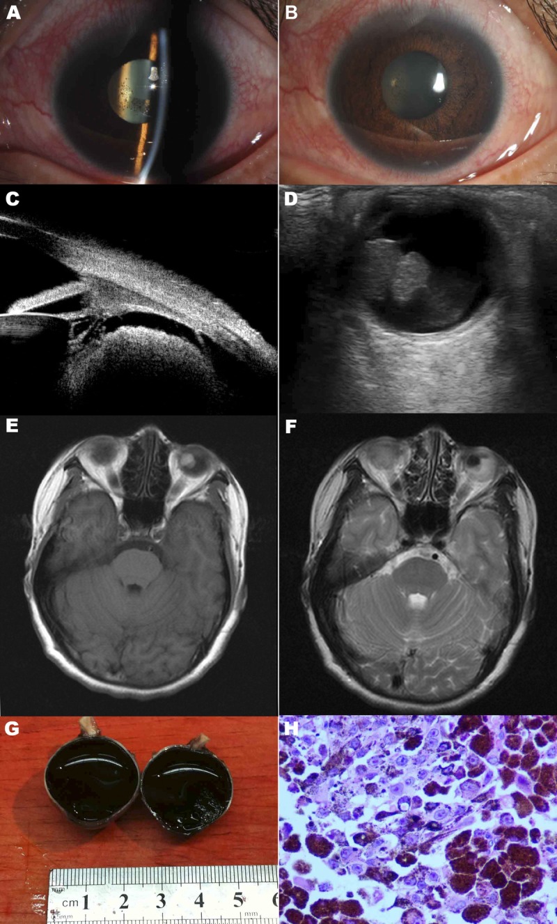
FIGURE 1.

Ophthalmologic findings of the left eye. (A, B) Slit lamp photography of the left eye showing ciliary injection, anterior chamber reaction, and keratic precipitates. (C) A tumor located behind the iris shown on UBM. (D) A large tumor in the peripheral choroid shown on B-scan ultrasonography. (E) T1-weighted contrast-enhanced magnetic resonance image of the brain and orbits showing a large mass in the left globe. (F) T2-weighted contrast-enhanced magnetic resonance image of the brain and orbits showing a large mass in the left globe. (G) Gross appearance of the tumor. (H) Pathologic section showing large round tumor cells with an epithelioid appearance. The cells contained abundant pink cytoplasm and dusty melanin. Single prominent eosinophilic nucleoli were seen in some melanocytes.
