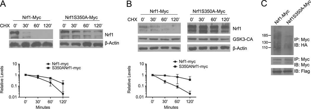
Fig. 5. GSK3 mediated degradation is attenuated by S350A substitution in Nrf1.
(A) HEK293 cells were transfected with Nrf1-myc or S350A–Nrf1-myc. Cells were then treated with 50 µg/ml cycloheximide, and harvested at the indicated time points for Western blotting using anti-Myc antibody. Beta-actin levels were used to determine loading of each sample. The graph shows quantitation of protein levels. Each point represents the mean±SEM of remaining protein. (B) HEK293 cells were co-transfected with Nrf1-myc, or S350A–Nrf1-myc and GSK3β-CA After 48 h, cells were treated with 50 µg/ml cycloheximide, and harvested at the indicated time points for Western blot analysis. Beta-actin levels were used to determine loading of each sample. Graph shows quantitation of the protein levels at each time points. Each point represents the mean±SEM of the remaining protein. (C) HEK293 cells were transfected with Nrf1-Myc or S350A–Nrfl-myc along with HA-ubiquitin and Flag-Fbw7. 48 h after transfections, cell extracts were prepared and immunoprecipitated with anti-Myc antibody, followed by immunoblotting with anti-HA antibody. For input control, the filter was stripped and probed with anti-Myc antibody. Cell extracts were also immunoblotted with anti-Flag antibody to determine Fbw7 expression.

