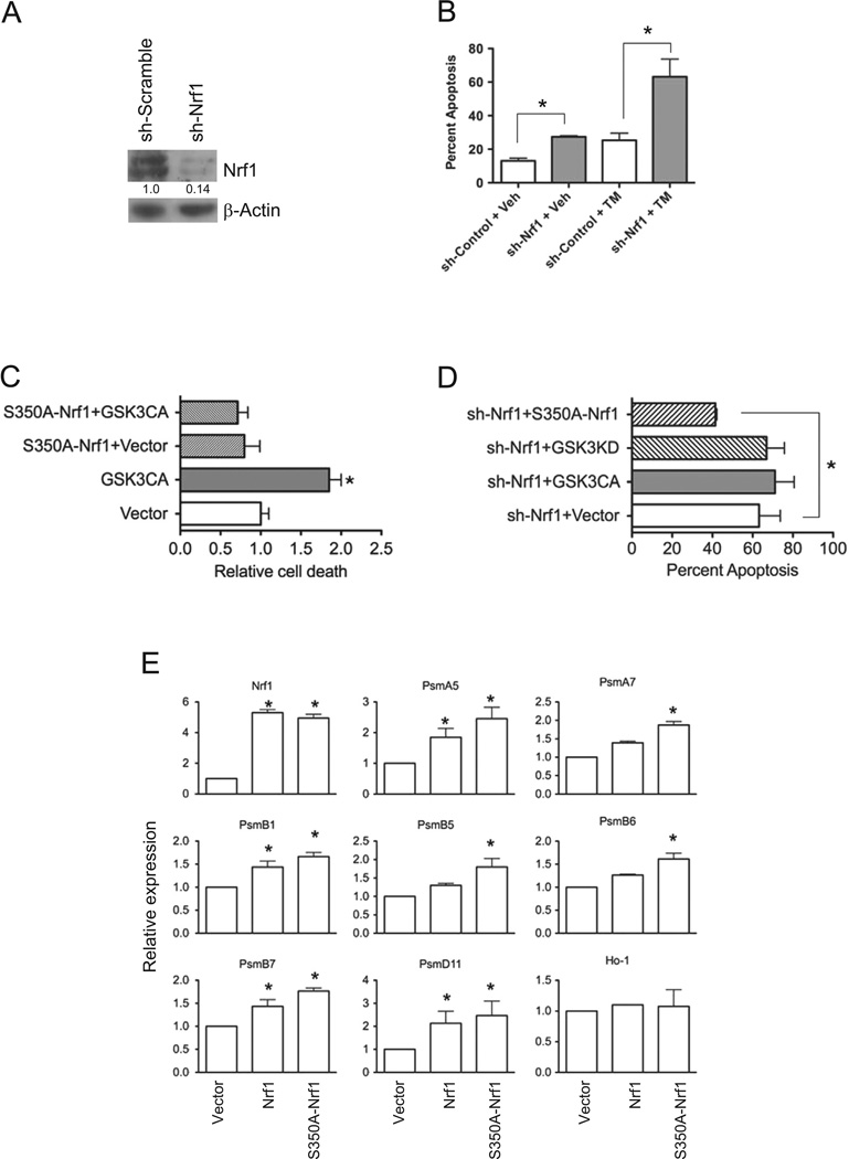
Fig. 6. GSK3 modulates Nrf1 mediated stress response in neuronal cells.
(A) Western blot analysis of Nrf1 in SH-SY5Y cells transduced with shScramble and shNrf1 virus. Densitometric quantitations of Nrf1 normalized against beta-Actin are shown. (B) shScramble- and shNrf1-transduced SH-SY5Y cells were treated with DMSO, or tunicamycin, and apoptosis was determined by Annexin V staining and flow cytometric analysis. Data represents means±SEM, n=3, *, P<0.05. (C) shScramble control cells co-transfected with EGFP and GSK3β-CA or EGFP, S350A–Nrf1 and GSK3β-CA were treated with tunicamycin, and cell death assessed by flow cytometric analysis of Annexin V staining in GFP-positive gated cells. Data represents means ± SEM, n=3, *, P<0.05. (D) shNrf1-transduced SH-SY5Y cells transfected with E-GFP and GSK3β-CA EGFP and GSK3β-KD, or EGFP and S350A–Nrfl were treated with tunicamycin. After 16 h, cell death assessed by flow cytometric analysis of Annexin V staining in GFP-positive gated cells. Data represents means±SEM, n=3, *, P<0.05. (E) Real time RTPCR analysis of gene expression in Nrf1 knockout MEF cells transfected with vector, Nrf1 or S350A–Nrf1. Expression levels of genes were quantitated relative to endogenous TBP levels as an internal reference, and calculated as 2(ct test gene-Ct TBP). Bars represent mean values±SEM of 3 experiments, and significance was assessed by Student’s t-test (*P<0.05).

