
Figure 1. Conditional deletion of NMHC II-A in the epidermis leads to epidermal hyperproliferation and disruption of hair follicle development in newborn mice.
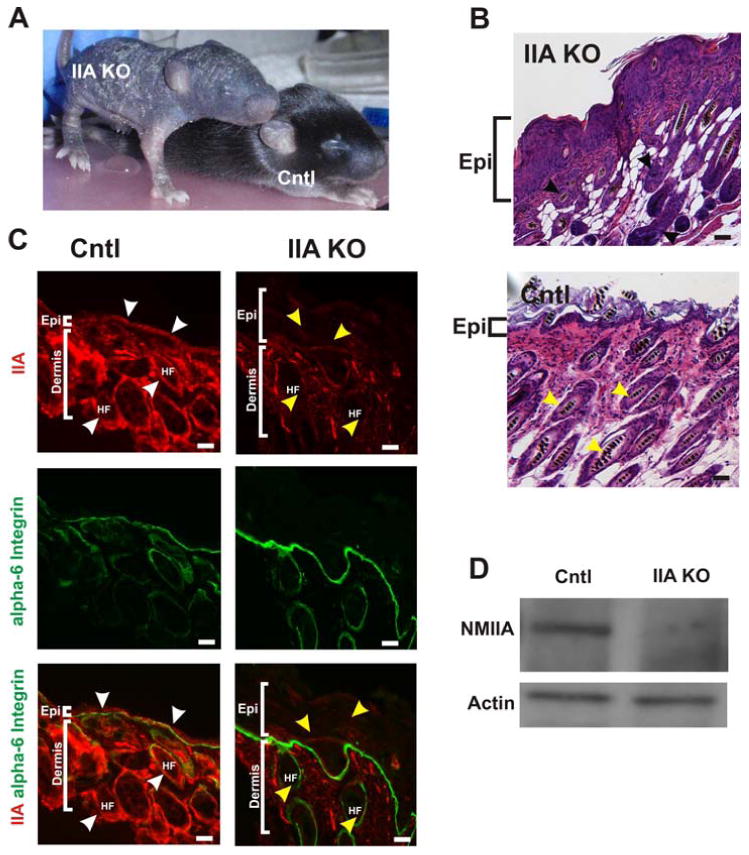
(A) The IIA KO mouse at postnatal day (P)10 displayed a severely disrupted skin and hair phenotype compared to the control littermate (Cntl) (B) Hematoxylin and Eosin (H & E) stained dorsal skin from P13 reveal a thickened and hyperproliferative epidermis (Epi) in the IIA KO (top panel) compared to the littermate epidermis (bottom panel) and disorganized and disoriented hair follicle growth in the IIA KO compared to control (black and yellow arrowheads, respectively). (C) Cryosections from the dorsal skin of both control and IIA KO P13 mice were immunostained for NM IIA (red) and alpha 6-integrin (green). White arrowheads point to NMIIA localization in the epidermis and hair follicle in the control epidermis. The yellow arrowheads identify representative regions in both the epidermis and hair follicles (HF) of the IIA KO that lack NM II expression. The thickened epidermis in the IIA KO is highlighted by the white brackets. Scale bars, 25μm. (D) Immunoblot analysis of P13 epidermal extracts from control and IIA KO mice and probed with anti-NMIIA antibody and actin which serves as a loading control.