
Figure 4. Epidermal loss of NMHC IIA drives suprabasal integrin ß1 activation.
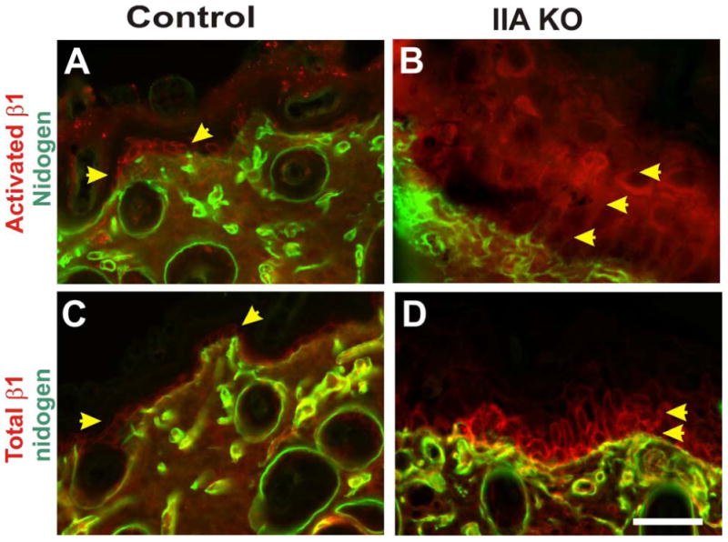
Skin from control littermate and IIA KO P13 mice was analyzed via immunohistochemistry on frozen sections. Control mouse skin was stained for activated integrin ß1 and nidogen, a basal lamina marker (A), or for total integrin ß1 and nidogen (C). In the IIA KO epidermis both activated integrin ß1 (B) and total integrin ß1 (D) display suprabasal localization. Yellow arrowheads denote single basal layer of the epidermis that expresses integrin ß1 in the control epidermis, and the expanded suprabasal expression observed in the IIA KO epidermis. Staining for activated integrin ß1 was performed using antibody clone 9EG7 (upper panels) and total integrin ß1 was performed using antibody clone MB1.2, as previously described (Honda et al, 2007). Scale bar, 50 μm.