
Figure 6. Placental defects are observed in the E12.5 IIA KO embryos.
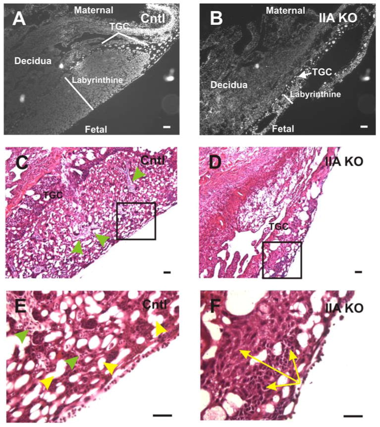
DAPI stained nuclei (gray scale) of control (A) and IIA KO (B) placentas. At E12.5, the IIA KO placenta exhibits a labyrinth whose area is dramatically reduced (white line) compared to the control placenta. The region of trophoblast giant cells (TGC) that normally surrounds the labyrinthine layer is thin and in some cases missing in the IIA KO placenta. H & E staining of control (C) and IIA KO (D) placentas reveals that the IIA KO placenta exhibits a lack of vascular infiltration originating from the maternal or fetal side, and a less porous network of sinusoidal spaces. The boxed regions in panels C and D are magnified in (E) and (F), respectively. The green arrowheads in (C) and (E) point to the well developed and organized trophoblast layer surrounding the vascular sinuses (yellow arrowheads, panel E) present in the control placenta. This ordered trophoblast cell-vascular sinus architecture is absent in the IIA KO placenta (F). Also, in panel F, the yellow arrows highlight a representative region of compacted trophoblast cells present throughout the labyrinthine layer of the IIA KO placenta but not evident in the control. Scale bars, 25 μm.