
Figure 9. Endogenous cytokeratin 5 expression in the E6.5 embryo is observed in the ectoplacental cone and trophectoderm regions in control embryos.
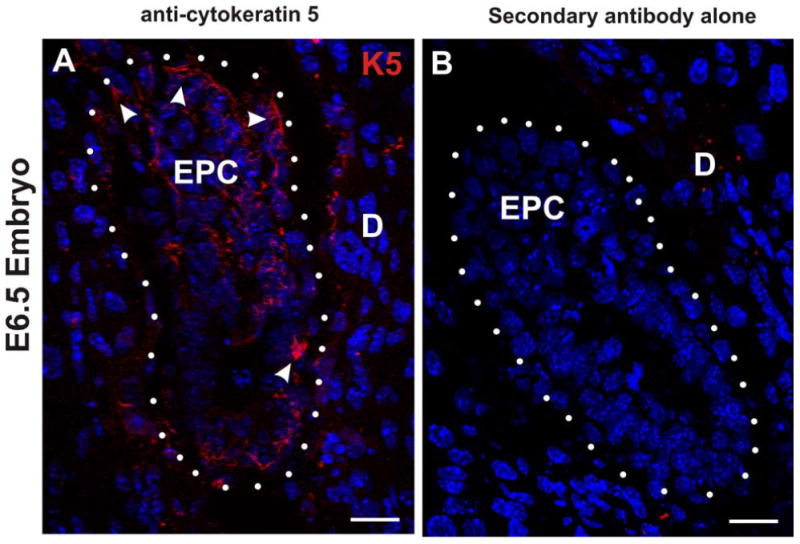
(A and B) Confocal immunofluorescence microscopy of frozen sections of E6.5 embryos. (A) Sagittal section of control E6.5 embryo was immunostained for cytokeratin 5 (K5; red) and stained with DAPI (blue). The white arrowheads identify K5 signal in the ectoplacental cone (EPC) and outer trophectoderm layer. The white dots enclose the embryo and denote the trophectoderm/decidua (D) border. (B) In the absence of primary antibody against cytokeratin 5 no immunostain signal is detected in the E6.5 embryo. Scale bars, 25 μm.