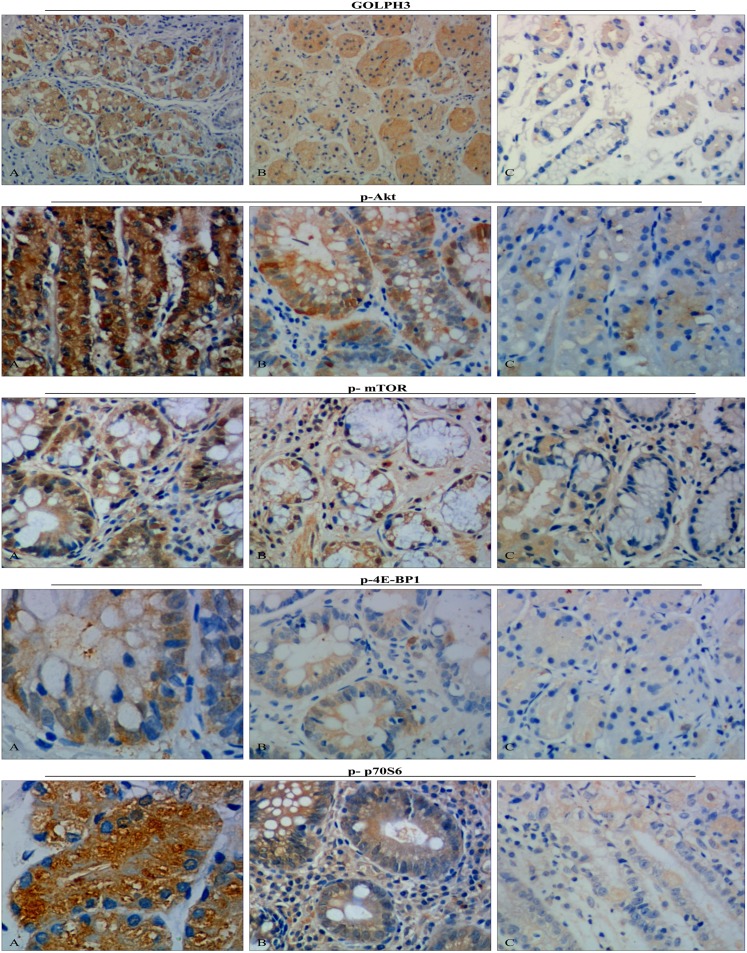
Figure 1. Representative GOLPH3, p-Akt, p-mTOR, p-4E-BP1 and p-p70S6 immunohistochemistry images in different tissues (brown granules, original magnification×400).
A: The gastric cancer group B: The carcinoma-adjacent tissue group C: The paired normal tissue group. Representative tissue sections from the gastric cancer group (n = 80), the carcinoma-adjacent tissue group (n = 80) and the paired normal tissue group (n = 80). Immunohistochemistry results demonstrated that the GOLPH3, p-Akt, p-mTOR and p-4E-BP1 were most expressed in the nucleus, the p-p70S6 was most expressed in the cytoplasm. The GOLPH3, phosphorylated Akt-mTOR Signaling pathway, p-4E-BP1 and p-p70S6 were highly expressed in gastric cancer groups.

