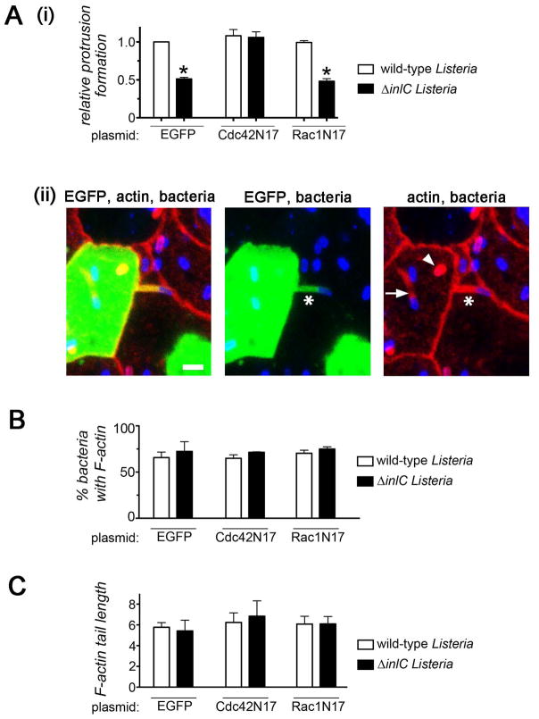
Figure 2. Inhibition of Cdc42 restores normal protrusion formation to Listeria lacking InlC.
A. Effect of Cdc42N17 on bacterial protrusion formation. Caco-2 BBE1 cells transiently expressing EGFP, EGFP-Cdc42N17, or EGFP-Rac1N17 were infected with wild-type or ΔinlC strains of Listeria for 5.5 hours, and assessed for protrusion formation or F-actin assembly as described in the Experimental Procedures. (A). Protrusion formation (i). Bar graph displaying mean relative protrusion formation values +/− SEM. (ii). Confocal microscopy images illustrating how protrusions were quantified. Protrusions (*) were identified as EGFP- positive F-actin tails projecting from transfected cells into neighboring EGFP-negative cells. Bacteria with F-actin tails (arrow) and symmetric F-actin (arrowhead) within the EGFP-positive cell body were also scored. Protrusion formation efficiency is expressed as the percentage of total bacteria-associated F-actin structures that were in protrusions. Relative protrusion efficiencies were calculated by normalizing absolute percentage values to that of the WT strain in EGFP-expressing cells. The scale bar indicates 5 micrometers. (B). Bacteria recruitment of F-actin. The mean percentage of bacteria +/− SEM that assembled F-actin in comet tails or symmetric structures is shown. (C) F-actin tail assembly. The bar graph contains mean F-actin tail lengths +/− SEM.

