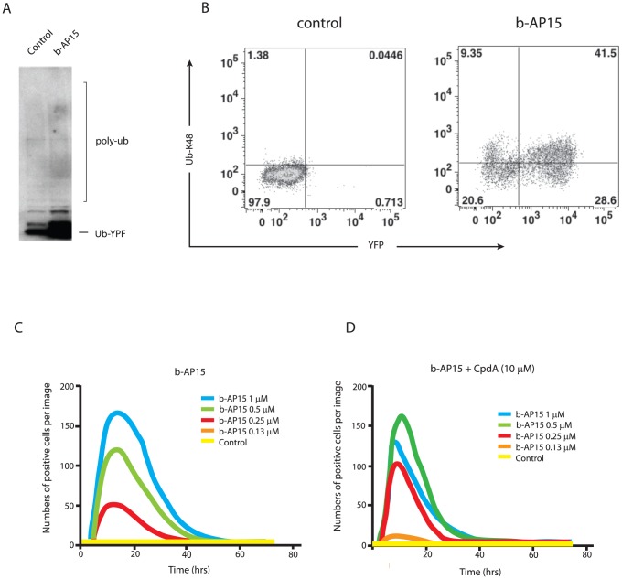
Figure 3. CpdA enhances the ability of b-AP15 to inhibit proteasomal degradation.
A. MelJuSo cells stably expressing UbG76V-YFP were treated with 1 µM b-AP15 for 6 hours or left untreated (control), and cell lysates subjected to immunoblotting using anti-YFP. The migration of UbG76V-YFP and the expected position of higher molecular weight polyubiquitinated forms (poly-Ub) are indicated. B. MelJuSo-UbG76V-YFP cells were exposed to 1 µM b-AP15 for 8 hours or left untreated (control). Cells were labeled with an anti-K48 polyubiquitin antibody followed by an allophycocyanin conjugated secondary antibody and analyzed by FACS. C. and D. MelJuSo-UbG76V-YFP cells were exposed to different concentrations of b-AP15 in the presence or absence of 10 µM cpdA as indicated. Changes in the number of fluorescence-positive cells/field following addition of the compounds were monitored using an IncuCyte-FLR microscope.

