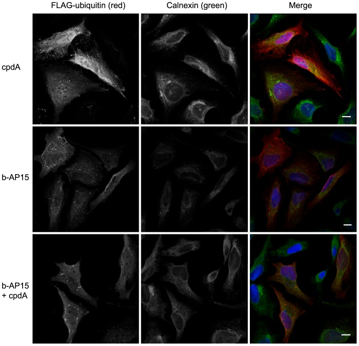
Figure 4. b-AP15 treatment alters the subcellular distribution of ubiquitin and causes the appearance of ubiquitin positive inclusions.
HeLa-M cells transiently transfected with FLAG-ubiquitin were treated with 10 µM cpdA (top panels), 1.0 µM b-AP15 (middle panels) or 0.8 µM b-AP15 plus 10 µM cpdA (bottom panels) for 16 h. Cells were fixed and stained with anti-FLAG and anti-calnexin antibodies followed by fluorescently labelled secondary antibodies. Confocal images were collected and images show the combined optical stacks. Scale bar = 10 µM.

